Overview of the Internal Anatomy of the Heart Anterior Dissection
The heart below is marked to show you where the two incisions should be made. Click on the tags below to find other quizzes on the same subject.
Solved Art Labeling Activity Overview Of The Internal Chegg Com
Identify the internal features of the heart.

. Use pointed scissors to open the right atrium. The skin overlying the thorax is innervated by the anterior and lateral cutaneous branches of the intercostal nerves ventral. The primary surface landmarks of the thorax are associated with the sternum jugular suprasternal notch sternoclavicular joint manubrium sternal angle and body clavicle ribs 2nd and 5th intercostal spaces nipple and mid-clavicular line.
Dissection Guide for Human Anatomy Lab 6 Objectives. Anatomy and Physiology questions and answers. The visceral layer of the serous pericardium aka epicardium is the outer layer of the heart itself.
A heart dissection is a fascinating experience that is at the core of our circulatory system and our body as a whole. You are basically going to be cutting each side of the heart so that you can look inside. Cardiovascular Human Dissection.
Note the general shape and size color and texture of the heart lungs and liver. 2 Remove the anterior thoracic wall. Optionally you may cut the heart in half to expose the chambers.
Where is it located compared to the trachea. The parietal layer of the serous pericardium is fused to the inside of the fibrous pericardium. Anterior interventricular coronary artery.
This layer is laden with fat in most hearts. The heart is surrounded by a serous sac. Follow the instructions on the guide to dissect a sheep heart.
External and internal anatomy aid in your understanding ofevollttion. Who are the experts. Made by Andrew Learn with flashcards games and more for free.
Experts are tested by Chegg as specialists in their subject area. Overview of the external anatomy of the heart anterior view Res Great cardiac vein Aortic arch Right coronary artery Left coronary artery Left pulmonary veins Ascending aorta Left pulmonary artery Anterior interventricular artery Superior vena cava Pulmonary trunk Auricle of. Even though this dissection has just a few cuts and some cleaning the anatomy of the heart may be challenging.
There our other dissection photos out there but I wanted to make a clear walkthrough for teachers and students who are doing it. Subdivisions of mediastinum 2. Heart Dissection External Questions 1 Label the heart view as anterior or posterior explaining what the answer means below 2.
4 Remove the right lung. Identify the internal structure of the heart and the arrangement of the four chambers. This quiz has tags.
Heart Anatomy internal parts PICS. Follow your TA to the lab where you will find a dissection guide. Quiz game to assist ap students with anatomy of exterior heart.
Mediastinum region central region of the thoracic cavity between the lungs and behind the. At least one blood vessel attaches. 3 Open the pleural sacs and define the pleural cavity parietal pleura and visceral pleura.
Coronary circulation anterior view The heart is actually one two or three pumps. Inspect the pleural sacs and mediastinum. JUN 28 74This is part of the OpenMichigan collection athttpopenumichedueducation.
To get a sense of the relationship of the heart and the pericardium in the body you can look at Video 519 from Aclands Video Atlas of Anatomy the link opens in a new tab. With this dissection a little bit of prep will go a long way. Middle Mediastinum Heart.
Some dissections will ask you to make a coronal cut where a single cut opens the entire back side of the heart. It is located in the. Return to the superior vena cava and the inferior vena cava.
The anatomy of the heart using a human cadaver. The aortic arch receive blood from the left ventricle of heart and reaches to all body partsThe vena ca. External anatomy of the heart anterior surface Art-labeling Activity.
Heart and Middle Mediastinum Reading. Anatomy of the heart 3. Art label different parts of human body.
G 354AN 217Gl 911 Use your forceps to remove as much clotted blood as possible from the SVC IVC and right atrium. View External_Heart_Dissection from BIO 234 at San Jose City College. We review their content and use your feedback to keep the quality high.
Start studying BIO2341-181 Gross Anatomy of the Heart Art-Labeling Activity. 1 Clean the thoracic body wall to demonstrate the sternum ribs costal cartilages and intercostal spaces. The following video is a step by step dissection of the heart and middle mediastinum.
Overview of the external anatomy of the heart. Draw an overview diagram in the ventral position lungs back with heart up at the front 3. The heart is a cone-shaped muscular organ about the size of your fist.
Anatomy and Physiology Art-Labeling Activity. Grays Anatomy for Students chapter 3 2. Physiology and behavior of fishes _ Working by yourself or with a team observe the external Of specimen complete the steps Of the dissection and identify as many as Written and visual elements Of the assigned handout.
The heart composed of two upper chamber -left and right atrium and two lower chamber -left and right ventricle. Aorta Left pulmonary artery Left atrium Mitral. Learn vocabulary terms and more with flashcards games and other study tools.
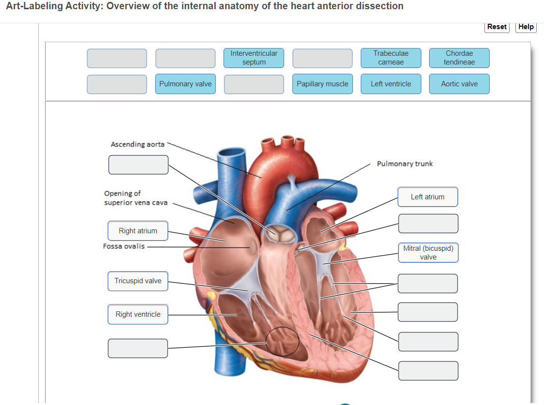
Art-labeling activityoverview of the internal anatomy of the heart anterior dissection. The picture below shows an anterior view of the heart with the pericardium removed. Overview of the internal anatomy of the heart anterior dissection Reset Help Interventricular septum Trabeculae carneae Chordae tendineae Pulmonary valve Papillary muscle Left ventricle Aortic valve Ascending aorta Pulmonary trunk Opening of superior vena cava Left atrium Right atrium Fossa ovalis Mitral bicuspid valve.
Circulation of the heart Clinical Correlates. Which chamber receives blood from the superior and inferior vena cavae. Heart Anatomy W pictures.
28 2016 by user Dongho Kim Heart Anatomy. To each of the chambers. Surface anatomy of the heart 3.
Coronary artery disease and associated. View the full answer. Heart Anatomy Created Nov.
Locate the following and identify the following in your drawing Esophagus this is the food tube. Start studying anatomy of the heart w pictures. The heart is a hollow organ containing 4 chambers.
Learn vocabulary terms and more with flashcards games and other study tools.

Anatomy Of Heart Interior Frontal Section Photos Prints Framed Puzzles 13014471

Human Heart Anatomy Poster Etsy Heart Anatomy Medical Anatomy Human Heart Anatomy

Anatomy Of The Heart Human Heart Anatomy Gross Anatomy Human Anatomy And Physiology

Chapter 19 The Circulatory System The Heart Ppt Download
Image Result For Gorgeous Facial Anatomy Illustrations Neuroscience Anatomy Art Medical Drawings Heart Anatomy

Heart Anatomy Medical Knowledge Anatomy

Images For Labeled Diagram Of The Heart Human Anatomy Picture Human Heart Anatomy Cardiac Anatomy

Pin By Scarlet On Medical Education Heart Structure Human Heart Sinusitis

Pin By Natasja Mitchell On School Female Anatomy Anatomy Atlas Anatomy

How The Human Heart Evolved Four Chambers Circulatory System Heart Structure Heart Anatomy

Pin On Biology Atlas

Internal Structure Of The Heart Contemporary Health Issues

Pin By Emily On Cardiovascular Icu In 2021 Heart Anatomy Human Heart Anatomy Medical Anatomy

Chapter 20 Cardiovascular System Flashcards Quizlet

The Heart Poster Allposters Com Heart Poster Neck Lump Behind Ear

Pin By Pnpueng On Anatomy Heart Anatomy Medical Knowledge Anatomy And Physiology

Heart 2 Anterior Labeled Anatomy Models Medical Anatomy Human Anatomy

The Internal Anatomy Of The Heart Anterior Dissection Diagram Quizlet

Trinx Human Heart Circulatory System Diagram Chart Medical Educational Science Class Anatomy Corazon Veins Arteries Labels White Wood Framed Art Poster 20x14 In 2022 Circulatory System Heart Structure Heart Anatomy
Comments
Post a Comment